[ad_1]
On August 23, doctors at the Churchill Hospital Oxford, in the U.K., conducted the country’s first uterus transplant. They removed the uterus from a 40-year-old woman and transplanted it to her 34-year-old sister, who had a rare condition that affected her ability to reproduce, according to the BBC. While the womb was functional, only a live birth in the near future can prove that the transplant succeeded, Isabel Quiroga, the lead surgeon involved in the procedure, has said.
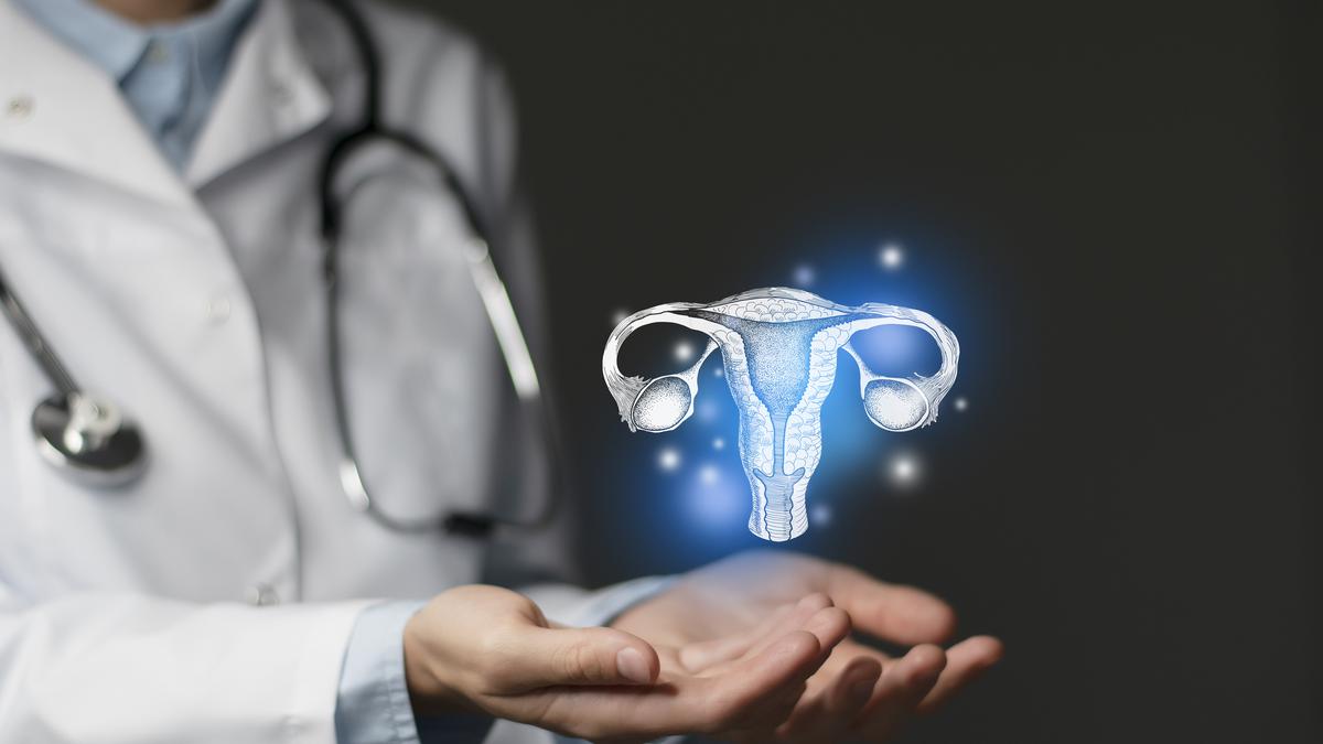
What is a uterus transplant?
Unlike heart or liver transplants, uterus transplants aren’t life-saving transplants. Instead, they are more like limb or skin transplants – which improve the quality of individuals’ lives. Uterus transplants can help women who lack a uterus fulfil their reproductive needs.
In 2014, as part of an initiative led by the chair of the obstetrics and gynaecology department at the University of Gothenburg, Mats Brännström, the first live birth after a uterus transplant took place in Sweden. The success signalled that the procedure could reverse the consequences of uterine factor infertility. Nine women in Sweden received transplanted wombs donated by relatives in Dr. Brännström’s programme, according to news reports.
The doctors now aim to make the procedure more affordable. At present, the National Health Service cost of surgery in the U.K. is estimated at GBP 25,000 (Rs 25.26 lakh).
India is one of a few countries to have had a successful uterine transplant; others include Turkey, Sweden, and the U.S. India’s first uterine transplant baby was born on October 18, 2018 – 17 months after the recipient had undergone the procedure. The cost of surgery is currently Rs 15-17 lakh in India.
What are the steps of a uterus transplant?
According to research papers published by the American Journal of Transplantation and the Journal of Clinical Medicine, before transplantation, the recipient is evaluated for good physical and mental health.
Similarly, whether the uterus is from a deceased or a live donor, it is checked for viability before it qualifies for donation. Live donors also undergo gynaecological examinations, including CT and MRI scans. They are also screened for uterine cancer, including tests for the human papillomavirus, a Papanicolaou smear, and an endometrial biopsy.
The procedure doesn’t connect the uterus to the fallopian tubes – which ensure the ovum from the ovaries moves to the uterus – so the individual can’t become pregnant through natural means. Instead, doctors remove the recipient’s ova, create embryos using in vitro fertilisation, and freeze them embryos (a.k.a. cryopreservation). Once the newly transplanted uterus is ‘ready’, the doctors implant the embryos in the uterus.
Once the transplant has been cleared, the uterus is carefully removed from the donor. The advent of robot-assisted laparoscopy has rendered the procedure less invasive.
The uterus is harvested together with its blood vessels. The arterial and venous vasculature (the network of vessels connecting the heart to other organs and tissues in the body), consisting of the deep uterine artery, the internal iliac arteries, the deep uterine vein, and the internal iliac veins are removed from either side. Surgeons also divide a part of the utero-ovarian branch to keep the ovarian veins from preserving the ovaries.
Surgeons also remove the fallopian tubes (and don’t use them in the graft for the donor to prevent pregnancies outside the uterus). With the recipient, the surgeons link up the muscles, cartilage, tendons, and arteries, veins, and other blood vessels so that the uterus functions normally.
What is a post-transplant pregnancy like?
Surgeons determine the transplant’s success in three stages.
The recipient’s chance of losing the graft is the highest in the first three months, so this is when doctors keep a tab on the graft’s viability.
Six months to one year after the procedure, doctors check for the proper function of the uterus. Regular menstruation is considered a good sign in this period. The recipient can attempt to conceive only after this phase.
In the first step of pregnancy, doctors transfer embryos prepared by in vitro fertilisation and cryopreserved to the recipient’s uterus. Just as with pregnancies after the transplants of other organs among women, there is a higher risk that the uterus will reject the uterus, or of spontaneous abortion, intrauterine death, low birthweight, or premature birth. So frequent check-ups and follow-ups are mandatory for women with transplanted uteri.
The final stage of success is of course successful childbirth.
Are there side-effects?
To prevent the recipient’s body from rejecting the transplanted uterus, the recipient needs to take drugs that suppress the immune system. These drugs are selected such that they won’t harm foetal development at any stage – from the uterus’s transplant until it is removed after childbirth.
These immunosuppressants are crucial but they do have other side-effects, including toxicity of the kidneys, bone-marrow toxicity, and a higher risk of developing diabetes and cancer. For these reasons, the uterus must be removed later.
The recipient is recommended regular follow-ups with doctors for at least a decade after the uterus’s removal to – among other things – keep on the lookout for potential long-term side effects of immunosuppressants.
Are there artificial uteri?
Successful uterus transplants have opened the door to new possibilities – including transplanting uteri from deceased donors, a process that has rarely succeeded. This could avoid the stigma and ethical concerns associated with utilising a live donor for a uterus transplant, which subjects a healthy person to medical procedures that can harm them for the benefit of another, especially when it’s not a life-saving condition, in contrast to deceased donation where the donor remains unharmed.
Dr. Brännström and his team at the University of Gothenburg are also working on creating a bioengineered artificial uterus. Such an entity could simplify the transplantation process by eliminating the need for live donors as well as sidestep debates about the ethics of using such organs.
According to a May 2017 paper authored by Dr. Brännström, to make a bioengineered uterus, researchers start with a small clump of stem cells taken from a woman’s blood or bone marrow and use it as the foundation for a 3D scaffold. New cells are added to this scaffold to build up a uterus. These experiments are still in their early stages; preliminary results with rats have shown some promise.
Dr. Brannstrom and his colleagues have estimated that it will be about a decade before artificial uteri will reach the efficiency and safety sufficient for human use.
When it does, the advantages could extend to women as well as members of the LGBTQ+ community. However, trans-women recipients, for example, will still need to undergo castration to create an artificial vagina – a process complicated by the fact that it involves hormones.
Male hormones, such as the androgens, can threaten a pregnancy, requiring the administration of high doses of counteracting exogenous hormones. Even then there are concerns about ensuring consistent blood flow to support a developing foetus, as the structure required for a uterus and for foetal development isn’t present in the male body.
[ad_2]

